Early-season stand loss of cotton could be the result of fungal pathogens or, in the case of some fields in the Upper Coast counties (e.g. Wharton and Fort Bend), high populations of reniform nematodes. Seedling death from fungi can vary from season to season and severe impacts are usually associated with cool, wet conditions at planting or shortly after emergence. Although the damage from stand loss can’t be undone during the current season, losses in future crops are somewhat preventable. In contrast, stand loss from the reniform nematode indicates a serious, long-term problem. In heavier soils, where the reniform nematode can thrive, it is essential to diagnose the cause of stand loss.
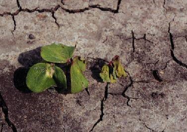
Figure 1. Post-emergence damping-off.
There are three major fungal pathogens causing seedling disease in Texas. The most prevelant pathogen is Rhizoctonia solani, which can cause seedling death after emergence (Figure 1). This fungus produces brown or black lesions on the stems. The lesions may be sunken, which is a symptom known as “soreshin” (Figure 2), or the lesions may girdle and pinch the stem at the soil surface, which is known as “wirestem”. Although this pathogen usually causes disease under cool, wet conditions, it can also cause disease at higher soil temperatures favorable for cotton seed germination, if cotton is planted into soil containing freshly-incorporated organic matter (e.g. a cover crop).
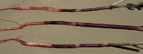
Figure 2. “Soreshin” symptoms caused by Rhizoctonia solani.
Several Pythium species cause seed decay and pre-emergent damping-off. The symptom on emerged seedlings is hypocotyl rotting below the soil surface. The hypocotyl has a water-soaked or light brown appearance. Pythium damping-off is usually associated with very wet soil.
Thielaviopsis basicola is a post-emergence pathogen that does not usually cause mortality, but may stunt plants and delay flowering. The disease it causes is known as “black root rot”. The fungus blackens the tap root and cortex (exterior) of the hypocotyl (Figure 3).
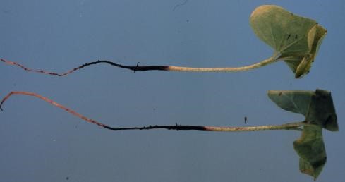
Figure 3. Symptoms of Thielaviopsis basicola.
Lateral roots are killed, particularly on older plants. This fungus is primarily a problem in the High Plains of Texas. It has been occasionally observed on plants from other growing areas of Texas.
Other fungal species have been reported to cause seedling disease in cotton, but their occurrence is infrequent and their impact relative to the three major pathogens is slight, unless they interact with the major pathogens. These pathogens tend to cause disease in plants growing under severe environmental stress. Fusarium species are prevalent, minor seedling pathogens, except for Fusarium oxysporum f. sp. vasinfectum race 4 (FOV4), which can be a significant seedling disease pathogen in fields located in El Paso and Hudspeth counties.
The reniform nematode can also cause stand loss (Figure 4). There are no obvious symptoms on plants early in the season, but symptoms may become more apparent later in the season, such as stunting and nutrient deficiency symptoms in the foliage.
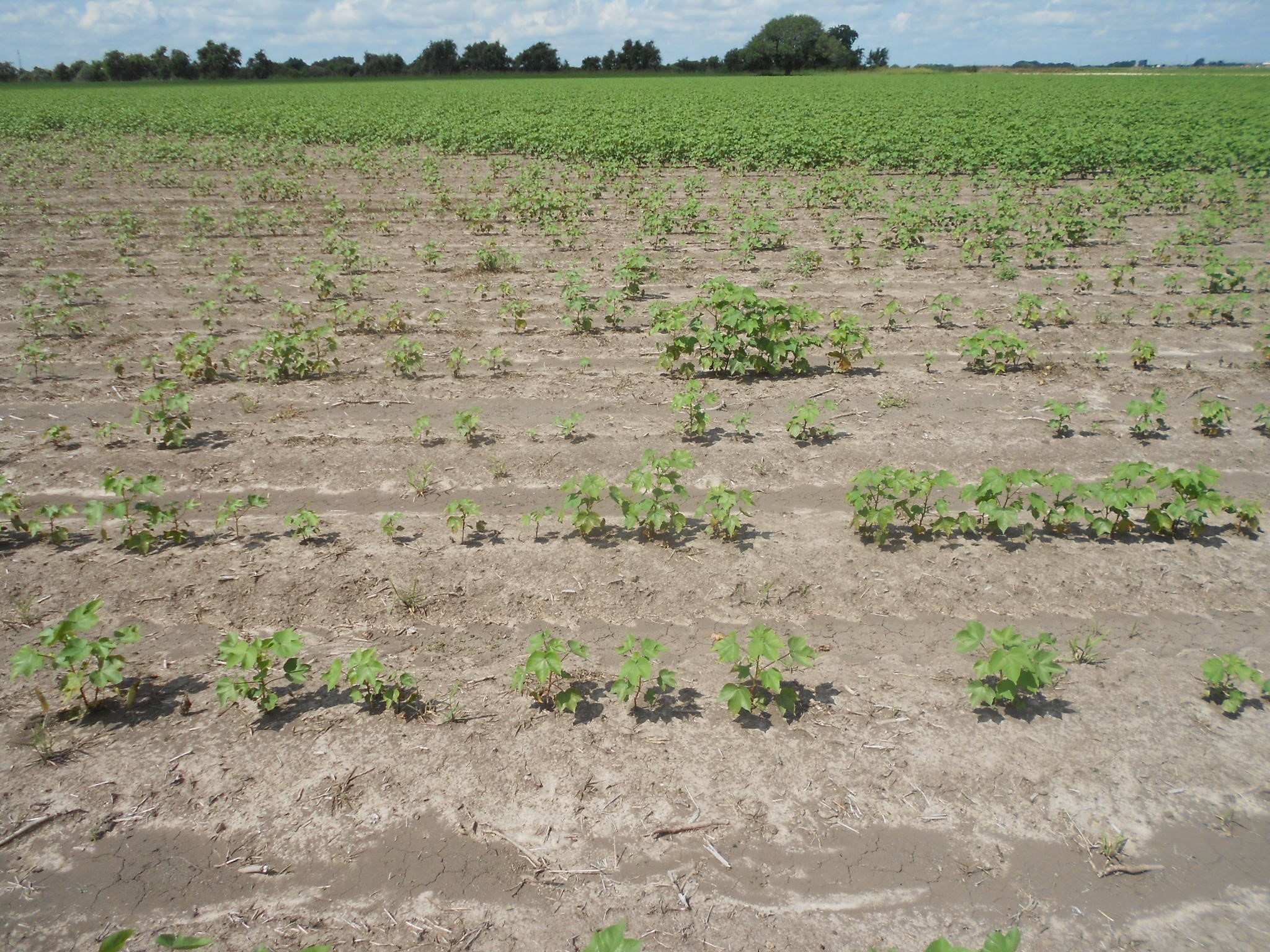
Figure 4. Stand loss caused by high populations of reniform nematode.
The reniform nematode can be diagnosed from soil samples. See https://agrilifecdn.tamu.edu/plantclinic/files/2010/10/D827r0627-FINAL.pdf for details on sampling and submission of samples to the Texas Plant Disease Diagnostic Clinic. If the nematode is present in a field, great care should be taken to prevent its movement to other fields via soil attached to implements. The nematode is very difficult to manage once it is present in a field and its presence could limit long-term productivity. In contrast, seedling diseases caused by fungi do not pose a long-term threat to productivity. See http://cotton.tamu.edu/Nematodes/Management%20of%20seedling%20diseases%20of%20cotton_2016.pdf for management guidelines.
Thomas Isakeit, Professor and extension plant pathologist
Texas A&M AgriLife Extension Service, College Station
t-isakeit@tamu.edu; cell 979-229-4976